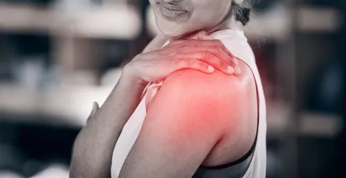
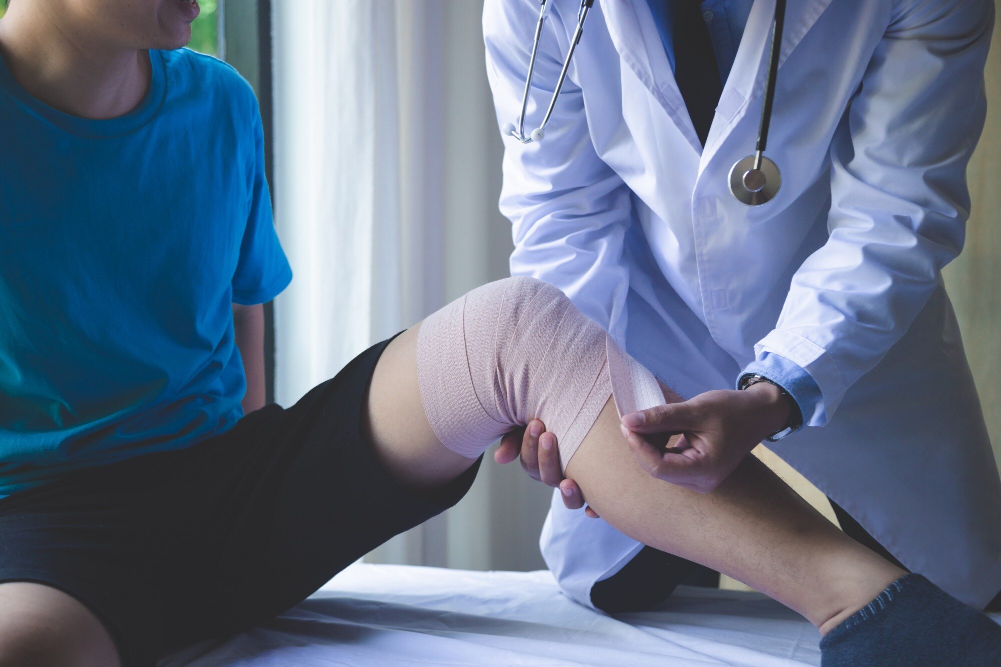
A dislocated joint needs more than a quick manoeuvre to put the bones back in place: clinicians must protect nearby nerves and blood vessels, identify fractures or trapped tissue, and support healing afterward. You will learn how to respond safely, what reduction and surgery involve, and which rehabilitation steps reduce the chance of another episode.
Key takeaways
- Immobilise the joint and seek urgent medical care; do not force it back into place.
- Clinicians use examination and imaging before reducing the joint safely.
- Surgery is considered for fractures, nerve or blood-vessel damage, and repeated instability.
- Strengthen the muscles controlling the joint and complete the prescribed rehabilitation plan.
How can you recognise a dislocation and protect the joint before care?
A dislocation occurs when the bones forming a joint are forced completely out of their normal alignment. Joint dislocation anatomy differs from a fracture, which is a broken bone; a sprain damages ligaments, a tendon injury affects muscle-to-bone tissue, and a partial dislocation or subluxation leaves some contact between the joint surfaces.
The joint’s appearance and symptoms vary. A shoulder dislocation can leave the arm held awkwardly; an elbow dislocation can cause marked deformity and inability to bend.
A finger may look crooked, a kneecap may shift sideways, a hip may cause severe pain with the leg shortened or turned, and a jaw dislocation can prevent the mouth from closing. Elbow dislocation emergency signs include numbness, a weak or absent pulse, or a pale, blue, cold hand.
Seek emergency care for any visible deformity, numbness, pale or blue skin, absent pulse, severe bleeding, an open injury, or suspected hip dislocation. Do not wait for swelling to settle.
- Keep the joint still in the position found. Do not straighten it or push it back into place; force can worsen a fracture or injure nerves, arteries, cartilage, and surrounding tissue.
- Remove rings, watches, or tight clothing before swelling increases.
- Apply a cold pack wrapped in cloth for 15–20 minutes at a time.
- Give no food or drink if sedation or surgery could be needed.
How do clinicians examine and realign a dislocated joint?
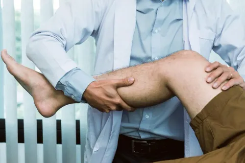
At the first assessment, clinicians inspect the deformity, swelling, wounds and skin, then palpate the joint and nearby bones. This dislocation physical examination identifies tenderness, instability and signs of an associated fracture.
A neurovascular assessment records skin colour and temperature, pulses and other blood-flow findings, sensation, and motor function below the injury, especially with shoulder, elbow, knee and hip dislocations.
1. The team compares movement and nerve function on both sides where safe. They document numbness, weakness, a pale or cold limb, and any absent pulse before treatment so a change after treatment is detectable.
2. An X-ray before and after reduction is commonly obtained. The first images look for a fracture or fracture-dislocation; the second confirms that the joint surfaces are aligned and identifies a new or previously hidden problem.
3. CT adds detail when X-rays do not define a complex fracture or bone defect. MRI can assess the labrum, ligaments, tendons and cartilage, while ultrasound can examine selected tendons, fluid collections or blood flow.
4. In a closed reduction, the clinician guides the joint surfaces into place without an incision, using a manoeuvre suited to the shoulder, elbow, knee or hip. Pain medicine, a local anaesthetic, closed reduction sedation, or general anaesthesia can reduce pain and muscle spasm.
Afterward, clinicians repeat the nerve, circulation, sensation and motor checks and often repeat imaging. A sling, splint or brace protects the realigned joint while damaged tissues are assessed.
When is surgery needed, and what does recovery after reduction involve?
Closed reduction is not enough when the joint cannot be safely realigned, a fracture-dislocation is present, tissue is trapped inside the joint, or a major nerve or blood-vessel injury needs repair.
Repeated instability that disrupts normal activity despite rehabilitation can also lead to open reduction or stabilisation surgery; a first dislocation does not automatically require an operation.
| Situation | Possible treatment | Why it matters |
|---|---|---|
| Irreducible joint | Open reduction | Muscle, bone, capsule, or other tissue blocks realignment |
| Fracture-dislocation | Fixation with stabilisation | A broken socket or bone can make the joint unstable |
| Trapped tissue or major neurovascular injury | Operative repair | The obstruction or damaged structure needs direct treatment |
| Repeated instability | Labral, capsular, or bone stabilisation | Repeated slipping can damage the joint further |
Hip dislocation treatment requires particular urgency because prolonged displacement can reduce blood flow to the femoral head and raise the risk of avascular necrosis. Associated fractures, unsafe closed reduction, or trapped tissue can require surgery.
After realignment, clinicians repeat circulation, sensation, and movement checks and often obtain follow-up imaging. A sling, splint, or brace supports early recovery while you follow restricted-movement instructions. Take prescribed pain medicine, keep any surgical wound clean and dry as directed, and attend every review.
Seek urgent reassessment for worsening pain, increasing swelling, numbness, weakness, a pale or cold limb, or a joint that slips out again. Immobilisation is temporary: once the joint is stable, clinician-directed motion must progress to strength and position-sense training, because prolonged immobilisation causes stiffness and muscle loss.
Why do dislocations recur, especially in the shoulder?
A successful reduction restores alignment; it does not repair the torn tissues, bone loss, or movement-control problem that caused instability. That is why a recurrent shoulder dislocation needs more than another period of immobilisation.
After a first traumatic shoulder dislocation, recurrence risk is shaped by:
- Age: adolescents and young adults face higher recurrence risk than older adults.
- Sport: contact, collision, throwing, and overhead sports repeatedly load the shoulder.
- Activity demands: work or recreation requiring forceful reaching, lifting, or extreme positions increases stress.
- Structural damage: a Bankart lesion involves the anterior labrum and capsule; a Hill-Sachs lesion is a compression defect in the humeral head; loss of glenoid bone reduces the socket’s containment.
A young athlete with a first traumatic dislocation may therefore discuss early stabilisation surgery, especially when imaging shows major labral or bone injury. An older adult with the same event may receive a different plan based on tissue quality, activity goals, stiffness risk, and associated rotator-cuff damage. Surgery is not automatic after every first dislocation.
Traumatic instability differs from multidirectional shoulder instability:
| Pattern | Main driver | Treatment emphasis |
|---|---|---|
| Traumatic | A discrete injury or bone defect | Imaging and structural stabilisation when indicated |
| Atraumatic or multidirectional | Weakness and poor neuromuscular control | Scapular, rotator-cuff, and shoulder-girdle rehabilitation first |
Recurrent episodes warrant X-rays and, when indicated, MRI or CT to assess the labrum, capsule, humeral head, and glenoid bone rather than repeating immobilisation alone.
Which rehabilitation steps reduce the chance of another dislocation?
To prevent repeated dislocations, complete rehabilitation in stages after the joint is stable: restore safe movement, build dynamic stabilisers, retrain balance and position sense, then practise joint-specific control before returning to sport or high-risk work.
- Begin with clinician-approved range-of-motion exercises. Forcing the joint too far can disrupt healing tissues.
- Add progressive resistance for the muscles that protect that joint, such as the rotator cuff and shoulder-blade muscles after a shoulder dislocation.
- Include proprioception exercises, which retrain your awareness of joint position through controlled balance, weight-shifting, and coordination drills.
- Rehearse sport or work movements at low speed before adding load, speed, contact, overhead reach, or uneven surfaces.
Pain disappearing is not clearance. Limited movement, weaker muscles, or poor control means the joint is not ready for return to sport after dislocation.
Treatment choice depends on the pattern:
| Option | What it usually involves | When it applies |
|---|---|---|
| Rehabilitation-first | Graduated mobility, strengthening, proprioception, and neuromuscular control | Atraumatic or multidirectional instability |
| Surgical assessment | Repair of the labrum or capsule, or restoration of damaged bone | Persistent function-limiting instability or a correctable structural defect |
An orthopaedic assessment at Dr DineshRam Orthopedician ECR can match the exercises and return-to-activity tests to your joint, imaging, and work or sport demands. Seek urgent reassessment if it slips again, or the limb becomes pale, cold, numb, weak, or increasingly swollen.
Related service
TRAUMA, FRACTURE & DISLOCATIONS TREATMENT Trauma, fractures, and dislocations can occur unexpectedly, often due to accidents, falls, or sports injuries, causing pain and temporarily affecting... View service → |
Frequently asked questions
How can you recognise a dislocation and protect the joint before care?
A dislocation can cause sudden severe pain, visible deformity, swelling, inability to move normally, numbness, or a cool or pale limb. Support the joint in its current position, remove rings or tight items, apply a wrapped cold pack, and seek urgent medical care. Do not try to force the joint back into place.
How do clinicians examine and realign a dislocated joint?
Clinicians check the joint’s shape, movement, skin colour, pulses, sensation, and nerve function. X-rays identify dislocation direction and fractures; other scans are used when soft-tissue damage needs assessment. After pain control or sedation, the clinician performs a reduction and repeats the neurovascular examination and imaging.
When is surgery needed, and what does recovery after reduction involve?
Surgery may be needed for an associated fracture, trapped tissue, nerve or blood-vessel injury, an irreducible dislocation, or repeated instability. Recovery commonly includes temporary immobilisation, pain control, gradual movement, and physiotherapy to restore range of motion and strength.
Why do dislocations recur, especially in the shoulder?
Recurrence can follow stretched or torn ligaments, a damaged labrum, bone loss, poor muscle control, or returning to sport before healing. The shoulder has a wide range of motion and relatively shallow socket, which makes it especially vulnerable to instability.
Which rehabilitation steps reduce the chance of another dislocation?
Follow the immobilisation period, attend physiotherapy, restore pain-free range of motion, and progressively strengthen the rotator cuff and shoulder-blade muscles. Practise joint-protection techniques and return to contact or overhead activities only after clinical clearance.